Texas A&M Agrilife Extension Service
Trending Searches:
Browse by Topic
Find research-based information and sustainable solutions created to improve the well-being of the land, people and animals across the State of Texas.


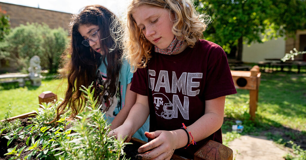
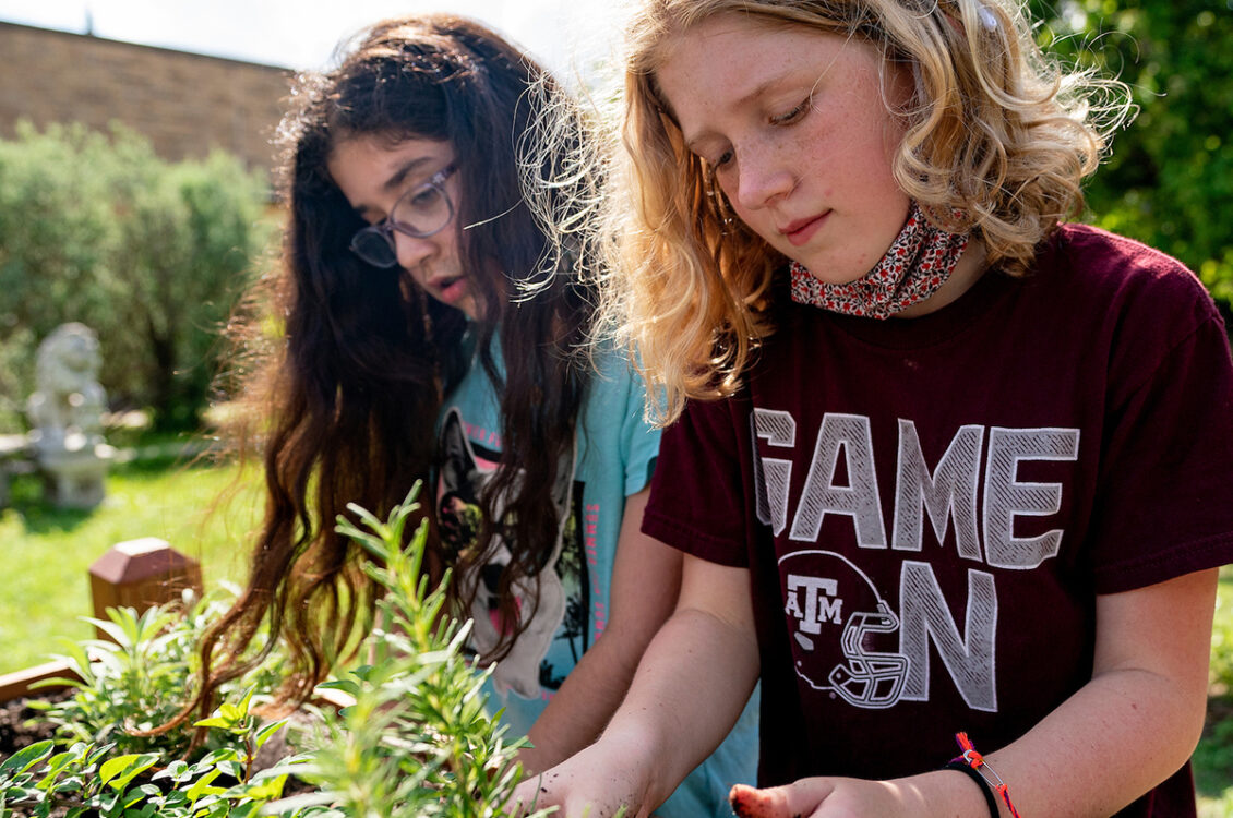
Texas 4-H Youth Development Program
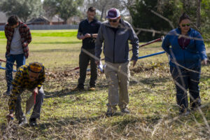
Texas 4-H is the largest youth development program in Texas reaching more than 550,000 youth each year. No matter where you live or what you like to do, Texas 4-H has something that lets you be a better you!
Find trustworthy solutions specific to your needs.
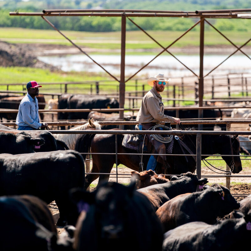
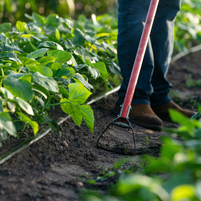
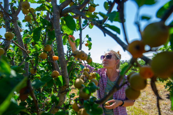
Scientific advances in agricultural and life sciences improve the well-being of Texas people, lands and businesses in meaningful and impactful ways. Find out how to bring these advances to your area of Texas.
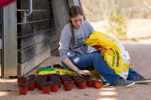

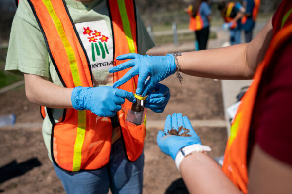
Give back to your community with AgriLife Extension-sponsored volunteer programs and opportunities in your area.
Get answers from our statewide network of experts through programs, testing services, tools and apps.

Grow your knowledge with a variety of research-based publications, self-paced courses, in-person workshops and more.
Find an AgriLife Extension representative in your area to receive personal support specific to your needs.
Three reasons to volunteer with the Texas A&M AgriLife Extension Service
Events